
DLAR Imaging Core (DLARIC)
The newly established Department of Laboratory Animal Resources Imaging Core (DLARIC), directed by Dr. Beth Bauer and managed by Betre Legesse, is located in the A. James Clark Hall vivarium. Technical and administrative support to the core is provided by Jared Robinson, Imaging Core Technical Coordinator. Raymond Chang provides technical consultation for the Albira PET-SPECT-CT imaging system. This core facility will provide imaging expertise, training and support for small animal- and cell/tissue-based research. The Core will offer imaging and irradiation services to all investigators at the University of Maryland. The DLARIC participates in the Olympus Discovery Center Program. This partnership provides researchers a great opportunity to tap into free, expert-led training and advanced consultation on two-photon microscopy.
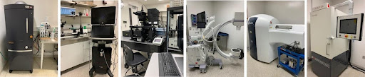
DLARIC resources include an array of cutting-edge in vivo, ex vivo and in vitro cellular, tissue and animal imaging modalities. Bioimaging instruments and other technologies include:
- IVIS Spectrum (for in vivo bioluminescence and fluorescence imaging that combines 2D and 3D optical tomography in a single platform);
- Visualsonics VEVO 3100 micro-ultrasound (ultra high-frequency ultrasound combining imaging and quantification in one touchscreen platform);
- Olympus multiphoton microscope FVMPE-RS (two-photon excitation fluorescent microscope which enables high-precision imaging of biological specimens to a depth of up to 1mm in living tissue and 3mm in fixed transparent specimens);
- GE Healthcare OEC One Mobile C-arm (fluoroscopic high-resolution X-ray images in real time);
- Bruker Albira PET-SPECT-CT (the trimodal integrative PET/SPECT/CT imaging allows quantitative 3D tomographic imaging of radiotracers, bone, and soft tissue); and a
- Precision X-Rad 320 (X-ray irradiator for cells, tissue and small animals up to rabbits).
Also see: DLARIC Imaging Equipment (PDF)
As part of the Olympus Discovery Center, Olympus is partnering with the University of Maryland to provide expert support and training on the FVMPE-RS microscope and to assist with new applications for optimal results.
Additionally, a dedicated surgical suite is available for imaging studies requiring intraoperative procedures. Trained users may register to reserve imaging facilities and equipment for use during normal business hours (8:00 AM to 4:30 PM Monday – Friday).
Fee Schedule:
Rates have been established for the following DLARIC services and equipment. (For additional rates, please visit the DLAR per diem and Animal Care/Support Rates page.)
Irradiation Services Rates (Precision X-Rad 320)
| Service | Rate | Unit |
|---|---|---|
| Tissues or Cells | $ 60.00 | run |
| Live Animals | $ 120.00 | run |
Bioimaging Equipment Rates
| Equipment | Rate | Unit |
|---|---|---|
| IVIS Spectrum | $ 60.00 | hour |
| VEVO 3100 micro-ultrasound | $ 60.00 | hour |
| Olympus multiphoton microscope FVMPE-RS | $ 105.00 | hour |
| OEC One Mobile C-arm | TBD | hour |
| Albira PET-SPECT-CT | TBD | hour |
| Equipment Training | $ 120.00 | hour |
Further information on available imaging Core services and resources may be found on the DLARIC website or by contacting the Core at DLARIC@umd.edu.