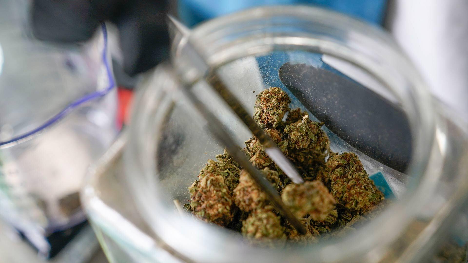
The U.S. House of Representatives’ vote on Friday to legalize cannabis exemplifies the growing acceptance of the drug both for medical and recreational use, but much remains to be understood about how it affects health—including how it might impact the cardiovascular system when smoked.
To help fill in that knowledge gap, a University of Maryland researcher is partnering with physicians at the University of Maryland School of Medicine (UMSOM) in a bid to use machine learning and advanced computing to obtain hitherto elusive answers about the hidden links between cannabis and heart health.
“We aim to determine whether there are morphological changes, changes in the structure or configuration of the heart chamber, or clinical values that are modified because of cannabis use,” said Eleonora Tubaldi, assistant professor of mechanical engineering. She is working with Dr. Jean Judy, a diagnostic radiology expert, and Dr. Timm Michael Dickfeld, a cardiac electrophysiologist—both professors at UMSOM.
The research is supported by an an MPower seed grant awarded by the University of Maryland Strategic Partnership: MPowering the State, which leverages the complementary strengths of University of Maryland, Baltimore (UMB) and the University of Maryland, College Park (UMCP).
To obtain their findings, the three will use the UK Biobank, a massive database that includes more than 40,000 images. Tubaldi will develop algorithms and a machine learning framework that will allow a computer to quickly and accurately compare MRI views of the cardiovascular systems of cannabis smokers to those of non-users. (Alternative methods of cannabis intake, such as edibles, are not part of the study.)
Machine learning can often detect changes that are too subtle to be registered by a human observer, Tubaldi said.
“We have limited vision and can see only some aspects of an image,” she said. “Machine learning can tell us about the texture of the image, or about contrast features that even a clinically trained eye might not be able to detect. It’s like a super-eye.”
It can also provide more consistent findings. “If you put ten radiologists in the same room, looking at the same image, they may very well not agree,” she said. “But with machine learning, instead of a person making a guess, we have a machine with an algorithm that knows precisely what to look for and can make a very precise observation.”
The team’s focus is on images obtained through a type of MRI known as T1 mapping, which tracks the relaxation rate of a certain tissue over a set time period. If the approach is successful, the scope may ultimately expand to include other types of imaging.
“There is a strong need to study this topic, and to be able to determine how much cannabis can be tolerated by the body,” Tubaldi said. “Such research can assist medical professionals and also inform policymakers.”
Original news story written by Robert Herschbach
